Atrial paced rhythm
A narrow vertical pacer spike immediately precedes each P wave: the pacemaker is starting the beat in the atria, and the impulse then conducts down the normal pathway. Normal PR and a narrow QRS follow, so the only tell is the spike in front of every P.
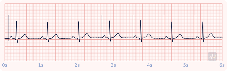
How to identify Atrial paced rhythm
- Rate
- The set pacing rate
- Rhythm
- Regular
- P waves
- Upright, one before every QRS, each right after a pacer spike
- PR interval
- 0.12-0.20 s, constant
- QRS
- Under 0.12 s
- Clincher
- A pacer spike immediately before every P wave, then a normal PR and a narrow QRS.
Rhythms it gets mistaken for
Atrial paced rhythm vs Ventricular paced rhythm
Atrial paced rhythm has a pacer spike right before every P wave. With a ventricular paced rhythm you would see a pacer spike right before every wide QRS. A narrow QRS means the signal came from above the ventricles.
Atrial paced rhythm vs Normal sinus rhythm
Atrial paced rhythm has a pacer spike right before every P wave. With normal sinus rhythm you would see no pacer spikes. A pacemaker starts every beat in the atria, and the impulse conducts down the normal pathway.
Atrial paced rhythm vs 1st-degree AV block
Atrial paced rhythm has a pacer spike right before every P wave. With 1st-degree block you would see no pacer spikes. A pacemaker starts every beat in the atria, and the impulse conducts down the normal pathway.
Treatment by certification level
Open your level. Treat the patient, not the strip.
Paramedic
Confirm the pacemaker is doing its job.
- Atrial capture: a P wave after every spike, then the patient's own narrow QRS through the normal pathway. Mechanical capture: a pulse to match.
- Failure to sense: spikes landing on the patient's own beats. Failure to capture: spikes with no P wave after them. Failure to pace: no spikes when there should be.
- If you defibrillate, keep the pad at least 1 inch from the implanted device, and never withhold a shock because of it.
AEMT
Pulse present and stable.
- Oxygen only if hypoxic or short of breath, IV access, monitoring.
- Acquire and transmit a 12-lead ECG. Interpreting it is a Paramedic skill.
- Transport and reassess. Request a paramedic if the patient worsens.
EMT
Pulse present: assess and support.
- Treat the patient, not the rhythm name: airway, breathing, circulation, vital signs.
- Oxygen if the SpO2 is under 94%, the patient is short of breath, or shows signs of shock.
- Request ALS and transport promptly. Reassess on the way.
An AED goes only on a patient who is unresponsive, not breathing and pulseless. It cannot feel a pulse, so on a patient with a pulse it can advise a shock that would do harm.