Rhythm Strip Library
All 32 rhythms on the Paramedic exam, from normal sinus rhythm through the AV blocks to the arrest rhythms. Each one has a clean 6-second example strip, the rate, rhythm, P waves, PR interval and QRS that identify it, the rhythms it gets mistaken for, and its treatment at the EMT, AEMT and Paramedic level.
Free, no account needed.
Sinus rhythms
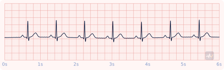 Normal sinus rhythmRate 60-100 and regular. One upright P before every QRS, PR 0.12-0.20 s, QRS under 0.12 s.
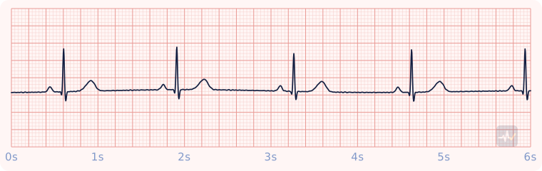 Sinus bradycardiaNormal P-QRS-T shape and regularity, just slow: rate under 60. Treat the patient, not the number. Only symptomatic bradycardia gets atropine or pacing.
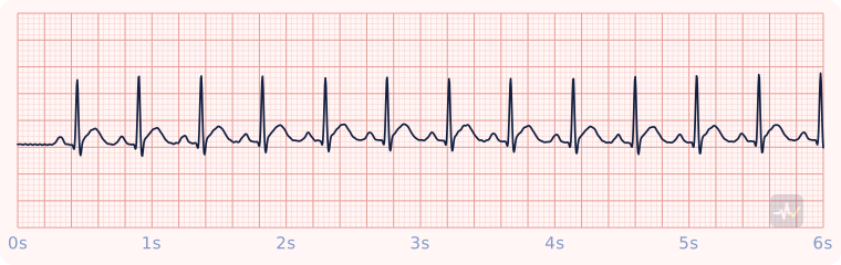 Sinus tachycardiaNormal P-QRS-T shape and regularity, just fast: rate over 100. It is almost always a response to something (pain, fever, hypovolemia, hypoxia, anxiety). Find and treat that cause rather than the rate.
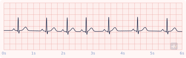 Sinus arrhythmiaEvery beat is a normal sinus beat, but the rate speeds up and slows down with breathing, so the R-R intervals stretch and shorten in a smooth, repeating wave.
Atrial rhythms
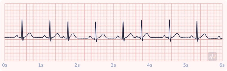 Premature atrial complexes (PACs)An otherwise sinus rhythm with beats that arrive early. Each early beat has a P wave that looks different from the sinus P (it can hide in the T wave before it) and a narrow QRS.
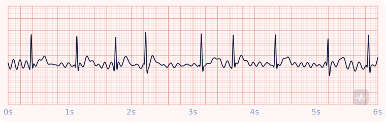 Atrial fibrillationIrregularly irregular R-R intervals with no discrete P waves, just a chaotic fibrillatory baseline. The QRS stays narrow because conduction below the AV node is normal.
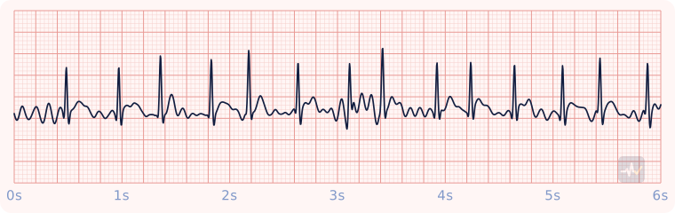 Atrial fibrillation with rapid ventricular responseThe same irregularly irregular, P-less rhythm as atrial fibrillation, but the ventricles are being driven fast, well over 100. The irregularity is harder to see at speed, so march out the R-R intervals.
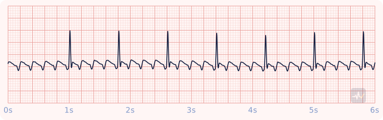 Atrial flutter (4:1)Sawtooth flutter waves at about 300/min. A fixed conduction ratio (here 4:1) gives a regular, much slower ventricular rate.
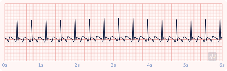 Atrial flutter (2:1)Flutter at 300 conducted 2:1 gives a regular narrow rhythm near 150. One flutter wave hides in each QRS or T, so a regular narrow tachycardia at almost exactly 150 earns a deliberate hunt for the sawtooth.
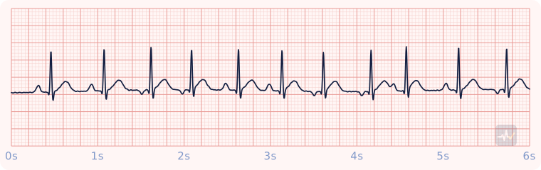 Multifocal atrial tachycardiaFast and irregular, with P waves of at least three different shapes and a PR interval that changes with them. Classically tied to severe COPD.
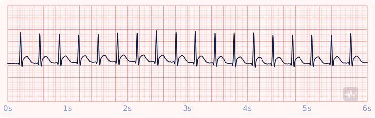 Supraventricular tachycardiaNarrow QRS, very fast (about 150-220) and metronome-regular, with P waves buried and not visible. The narrow QRS is what separates it from V-tach.
Junctional rhythms
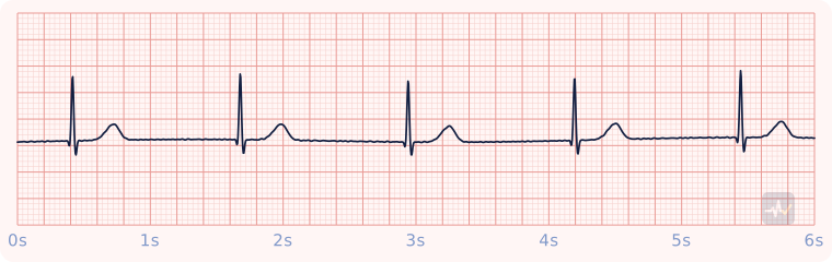 Junctional escape rhythmThe AV junction takes over at its own 40-60 when the sinus node fails. Narrow QRS, but no upright P before it: the P is absent, inverted, or hidden in the QRS.
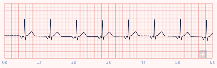 Accelerated junctional rhythmA junctional rhythm running 60-100. The impulse spreads backward into the atria, so the P wave is inverted and sits just in front of the narrow QRS with a short PR (or hides in or after it).
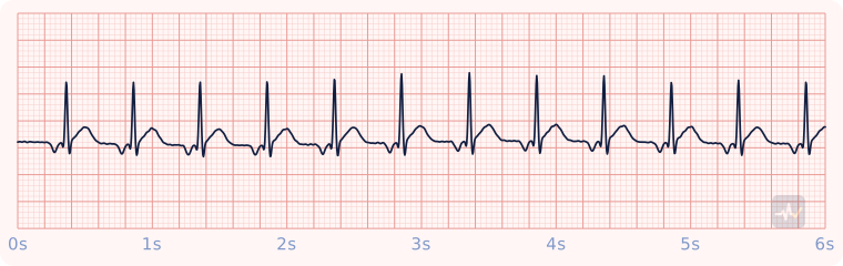 Junctional tachycardiaA junctional rhythm running faster than 100. Narrow and regular, with the P wave absent, inverted just in front of the QRS, or retrograde just after it. The rate over 100 separates it from accelerated junctional (60-100). The inverted P separates it from sinus tach, and from SVT, which usually runs faster with its P waves buried.
AV blocks
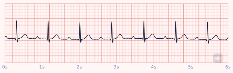 1st-degree AV blockEvery P conducts, but slowly: the PR interval is longer than 0.20 s and the same length on every beat. No QRS is ever dropped. A finding more than a rhythm.
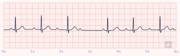 2nd-degree type I (Wenckebach)The PR interval lengthens beat after beat until one P wave is not conducted and its QRS drops, then the cycle restarts. Longer, longer, longer, drop. Usually nodal and usually benign.
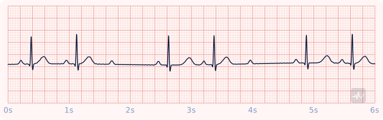 2nd-degree type II (Mobitz II)The PR on conducted beats is constant and never lengthens, yet P waves are intermittently not conducted and a QRS drops without warning. It sits below the AV node and can progress abruptly to complete block.
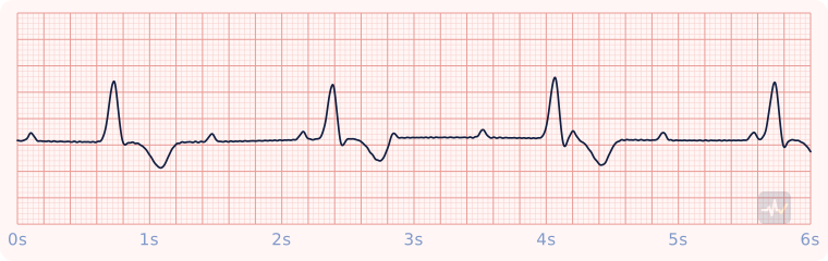 3rd-degree (complete) AV blockComplete AV dissociation. P waves march out at their own regular rate and wide escape complexes march out at a separate, slower rate, with no relationship between them. The PR is different on every beat.
Ventricular rhythms
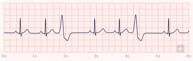 Unifocal PVCsAn otherwise sinus rhythm interrupted by early, wide, bizarre complexes with no P wave in front and a T wave pointing opposite the QRS. Every PVC has the same shape, and the pause after each one is fully compensatory.
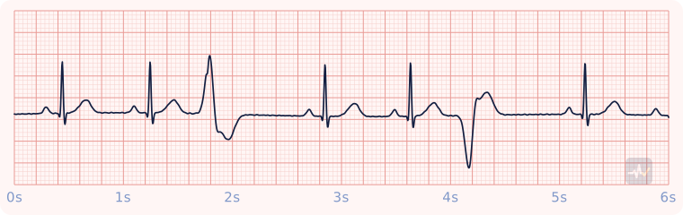 Multifocal PVCsPVCs that do not match each other: here one points up and one points down. Different shapes mean more than one irritable spot in the ventricle.
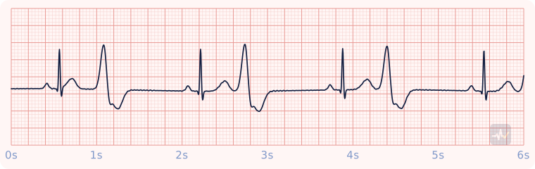 Ventricular bigeminyEvery other beat is a PVC: normal, PVC, pause, normal, PVC, pause. Each PVC is coupled to the sinus beat before it at the same interval.
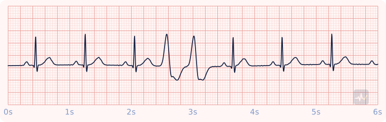 PVC coupletTwo PVCs in a row. Three or more in a row is a run of ventricular tachycardia.
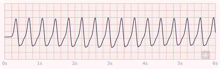 Ventricular tachycardia (monomorphic)Wide, bizarre QRS complexes (here about 0.20 s) at 150-250, regular, every complex the same shape, no identifiable P waves. A wide, fast, regular rhythm you cannot classify is V-tach until proven otherwise.
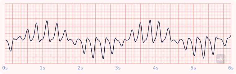 Torsades de pointesPolymorphic V-tach: wide, irregular complexes whose height and direction twist around the baseline in a spindle pattern. It grows out of a prolonged QT, and magnesium sulfate is the specific drug.
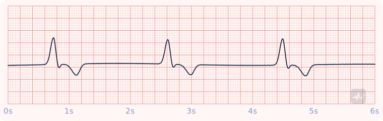 Idioventricular rhythmThe ventricle escaping on its own at 20-40. Very wide complexes, no P waves, very slow. It may be the only thing keeping the patient alive, so it is never suppressed with an antiarrhythmic.
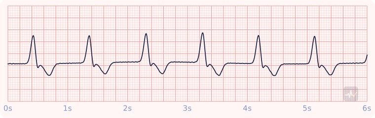 Accelerated idioventricular rhythm (AIVR)A ventricular rhythm running 40-100: wide, regular, no P waves, faster than an escape rhythm but slower than V-tach. Common after reperfusion.
Arrest rhythms
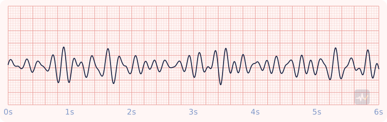 Ventricular fibrillation (coarse)Chaotic, disorganized waves with no P, QRS or T and no pulse. Coarse V-fib has large waves. Shockable: defibrillate and go straight back to compressions.
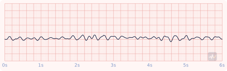 Ventricular fibrillation (fine)The same chaos at low amplitude. Fine V-fib can look almost flat, which is why a flat line is confirmed in a second lead before anyone calls it asystole.
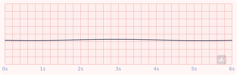 AsystoleA flat line with only slow baseline drift. Confirm it in a second lead and check leads, cables and gain before calling it. Not shockable: CPR and epinephrine.
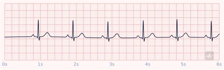 Pulseless electrical activity (PEA)Any organized rhythm with no pulse. This strip would be a normal rhythm if the patient had a pulse, which is the point: PEA is found with your fingers, not on the paper. Not shockable, it is a hunt for the cause.
Paced rhythms
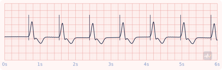 Ventricular paced rhythmA narrow vertical pacer spike immediately precedes each wide QRS: the pacemaker, not the AV node, is depolarizing the ventricle. Electrical capture is a wide complex after every spike; mechanical capture is a pulse to match.
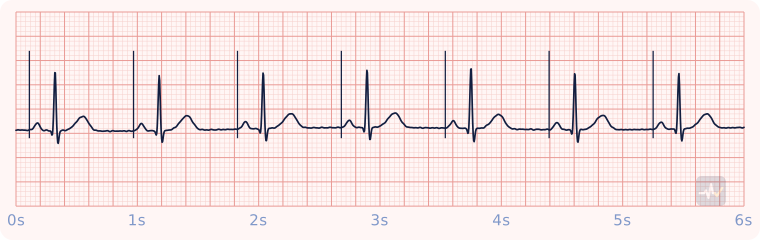 Atrial paced rhythmA narrow vertical pacer spike immediately precedes each P wave: the pacemaker is starting the beat in the atria, and the impulse then conducts down the normal pathway. Normal PR and a narrow QRS follow, so the only tell is the spike in front of every P.