Multifocal PVCs
PVCs that do not match each other: here one points up and one points down. Different shapes mean more than one irritable spot in the ventricle.
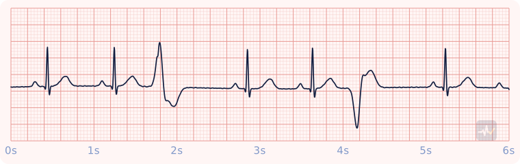
How to identify Multifocal PVCs
- Rate
- That of the underlying rhythm
- Rhythm
- Regular, interrupted by early beats
- P waves
- None in front of the early beats
- PR interval
- Normal on the sinus beats
- QRS
- Early beats are wide, and they differ from each other
- Clincher
- PVCs of more than one shape: more than one irritable spot.
Rhythms it gets mistaken for
Multifocal PVCs vs Unifocal PVCs
Multifocal PVCs has wide early beats of more than one shape. With unifocal PVCs you would see wide complexes that all look alike.
Multifocal PVCs vs PVC couplet
Multifocal PVCs has wide early beats of more than one shape. With a PVC couplet you would see wide complexes that all look alike.
Multifocal PVCs vs Ventricular bigeminy
Multifocal PVCs has wide early beats of more than one shape. With bigeminy you would see wide complexes that all look alike.
Treatment by certification level
Open your level. Treat the patient, not the strip.
Paramedic
Treat the cause and monitor.
- No prophylactic antiarrhythmic for isolated ectopy, even in acute coronary syndrome. Treat the ischemia and watch the rhythm.
- Watch for the patterns that matter: R-on-T, couplets, multiformed, frequent uniform, bigeminy.
AEMT
Pulse present and stable.
- Oxygen only if hypoxic or short of breath, IV access, monitoring.
- Acquire and transmit a 12-lead ECG. Interpreting it is a Paramedic skill.
- Transport and reassess. Request a paramedic if the patient worsens.
EMT
Pulse present: assess and support.
- Treat the patient, not the rhythm name: airway, breathing, circulation, vital signs.
- Oxygen if the SpO2 is under 94%, the patient is short of breath, or shows signs of shock.
- Request ALS and transport promptly. Reassess on the way.
An AED goes only on a patient who is unresponsive, not breathing and pulseless. It cannot feel a pulse, so on a patient with a pulse it can advise a shock that would do harm.