Ventricular fibrillation (fine)
Also called fine V-fib.
The same chaos at low amplitude. Fine V-fib can look almost flat, which is why a flat line is confirmed in a second lead before anyone calls it asystole.
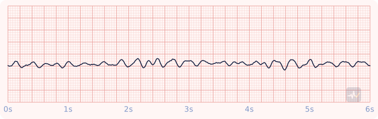
How to identify Ventricular fibrillation (fine)
- Rate
- None to count
- Rhythm
- Chaotic
- P waves
- None
- PR interval
- None
- QRS
- None, only low, disorganized waves
- Clincher
- Low-amplitude chaos that can pass for a flat line. Confirm a flat line in a second lead.
Rhythms it gets mistaken for
Ventricular fibrillation (fine) vs Asystole
Ventricular fibrillation (fine) has only chaotic waves, with no P, QRS or T. With asystole you would see a flat line. It starts in the ventricles: a ventricular rhythm.
Ventricular fibrillation (fine) vs Ventricular fibrillation (coarse)
Ventricular fibrillation (fine) has low chaotic waves that can pass for a flat line. With coarse V-fib you would see large chaotic waves.
Ventricular fibrillation (fine) vs Idioventricular rhythm
Ventricular fibrillation (fine) has only chaotic waves, with no P, QRS or T. With an idioventricular rhythm you would see distinct QRS complexes you can pick out.
Treatment by certification level
Open your level. Treat the patient, not the strip.
Paramedic
Shockable arrest.
- CPR, pads on. Defibrillate (unsynchronized): biphasic 120-200 J (the maximum if you do not know the device recommendation), monophasic 360 J.
- Resume compressions immediately after the shock. No rhythm or pulse check first.
- IV/IO access. Epinephrine 1 mg every 3-5 minutes, typically after the second shock.
- Still shockable after a shock and epinephrine: amiodarone 300 mg IV/IO, then 150 mg if it persists. Lidocaine 1-1.5 mg/kg, then half that, is the alternative.
- Two-minute cycles: rhythm check, shock if shockable. Waveform capnography throughout.
- Look for and treat the cause: the H's and T's.
AEMT
Cardiac arrest: CPR, the AED, epinephrine.
- High-quality CPR and the AED, exactly as at the EMT level. After every analysis, shock or no shock, compressions resume immediately.
- IV or IO access.
- Epinephrine 1 mg IV/IO every 3-5 minutes, typically started after the second shock.
- Supraglottic airway and waveform capnography. EtCO2 under 10 mmHg means fix the compressions; an abrupt sustained rise suggests ROSC, confirmed at the scheduled rhythm check.
Manual defibrillation and antiarrhythmics (amiodarone, lidocaine) are Paramedic level. The AEMT's electrical therapy is the AED.
EMT
Cardiac arrest: CPR and the AED.
- High-quality CPR, starting with compressions.
- AED on as soon as it arrives.
- The AED will advise a shock for this rhythm. Clear, shock, then resume compressions immediately without a pulse check.
- Two minutes of CPR, then let the AED reanalyze. Check a pulse only at that scheduled rhythm check.