3rd-degree (complete) AV block
Also called complete heart block, third-degree heart block.
Complete AV dissociation. P waves march out at their own regular rate and wide escape complexes march out at a separate, slower rate, with no relationship between them. The PR is different on every beat.
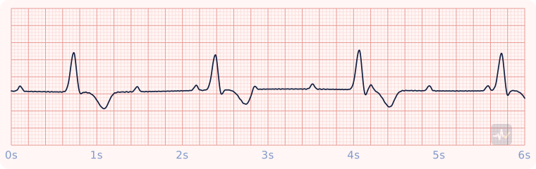
How to identify 3rd-degree (complete) AV block
- Rate
- Atrial faster than ventricular. The escape runs 40-60 if junctional, 20-40 if ventricular
- Rhythm
- P-P regular and R-R regular, independently
- P waves
- Regular, marching through the QRS and T waves
- PR interval
- Different on every beat: no relationship
- QRS
- Wide here (a ventricular escape)
- Clincher
- Two rhythms that ignore each other: AV dissociation.
Rhythms it gets mistaken for
3rd-degree (complete) AV block vs 2nd-degree type II (Mobitz II)
3rd-degree (complete) AV block has no relationship between the P waves and the QRS complexes. With Mobitz II you would see a PR interval that stays the same, then a QRS that drops without warning. A wide QRS means the signal started in the ventricles or got there through an abnormal path.
3rd-degree (complete) AV block vs Idioventricular rhythm
3rd-degree (complete) AV block has regular P waves, with more P waves than QRS complexes. With an idioventricular rhythm you would see no upright P wave in front of the QRS. It is an AV block: the P waves come from the SA node, and the problem is the P-to-QRS relationship.
3rd-degree (complete) AV block vs 2nd-degree type I (Wenckebach)
3rd-degree (complete) AV block has no relationship between the P waves and the QRS complexes. With Wenckebach you would see a PR interval that gets longer beat after beat until a QRS drops. A wide QRS means the signal started in the ventricles or got there through an abnormal path.
Treatment by certification level
Open your level. Treat the patient, not the strip.
Paramedic
Stable
Stable, but watch it closely.
- Monitor, IV access, transport and reassess.
- Put the pads on now so pacing is ready.
- If it becomes symptomatic, go to pacing or dopamine 5-20 mcg/kg/min, or epinephrine 2-10 mcg/min, titrated to effect. Do not rely on atropine: the block sits below where it acts.
Unstable
Symptomatic high-grade block: go to pacing.
- Do not rely on atropine: the block sits below where it acts. Atropine 1 mg may be tried while the pads go on, but it never delays pacing.
- Transcutaneous pacing (rate 60-80, mA up until capture, then slightly above). Confirm mechanical capture with a femoral pulse, not a carotid.
- Sedation and analgesia; recheck the blood pressure once captured.
- No capture or still poorly perfusing: dopamine 5-20 mcg/kg/min, or epinephrine 2-10 mcg/min, titrated to effect.
- Expert consultation: this patient may need a transvenous pacer.
AEMT
Stable
Pulse present and stable.
- Oxygen only if hypoxic or short of breath, IV access, monitoring.
- Acquire and transmit a 12-lead ECG. Interpreting it is a Paramedic skill.
- Transport and reassess. Request a paramedic if the patient worsens.
Unstable
Symptomatic bradycardia with a pulse.
- Support oxygenation and ventilation, establish IV access, and move the patient.
- Expedited transport, with a paramedic intercept if that is genuinely faster than reaching the hospital.
Synchronized cardioversion and pacing are Paramedic-only, so the AEMT has no electrical option for a patient with a pulse. Never put the AED on this patient.
EMT
Stable
Pulse present: assess and support.
- Treat the patient, not the rhythm name: airway, breathing, circulation, vital signs.
- Oxygen if the SpO2 is under 94%, the patient is short of breath, or shows signs of shock.
- Request ALS and transport promptly. Reassess on the way.
An AED goes only on a patient who is unresponsive, not breathing and pulseless. It cannot feel a pulse, so on a patient with a pulse it can advise a shock that would do harm.
Unstable
Pulse present but poorly perfusing: this patient needs ALS fast.
- Treat the patient, not the rhythm name: airway, breathing, circulation, vital signs.
- Oxygen if the SpO2 is under 94%, the patient is short of breath, or shows signs of shock.
- Request ALS and transport promptly. Reassess on the way.
An AED goes only on a patient who is unresponsive, not breathing and pulseless. It cannot feel a pulse, so on a patient with a pulse it can advise a shock that would do harm.