Torsades de pointes
Polymorphic V-tach: wide, irregular complexes whose height and direction twist around the baseline in a spindle pattern. It grows out of a prolonged QT, and magnesium sulfate is the specific drug.
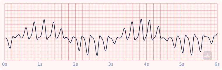
How to identify Torsades de pointes
- Rate
- Very fast
- Rhythm
- Irregular
- P waves
- None
- PR interval
- None
- QRS
- Wide, changing height and direction
- Clincher
- Complexes that twist around the baseline in a spindle pattern.
- Common causes
- A prolonged QT: QT-prolonging medications, electrolyte problems (low potassium), congenital long QT.
Rhythms it gets mistaken for
Torsades de pointes vs Ventricular tachycardia (monomorphic)
Torsades de pointes has complexes that change height and direction, twisting around the baseline. With V-tach you would see wide complexes that all look alike.
Torsades de pointes vs Ventricular fibrillation (coarse)
Torsades de pointes has distinct QRS complexes you can pick out. With coarse V-fib you would see only chaotic waves, with no P, QRS or T.
Torsades de pointes vs Ventricular fibrillation (fine)
Torsades de pointes has distinct QRS complexes you can pick out. With fine V-fib you would see only chaotic waves, with no P, QRS or T.
Treatment by certification level
Open your level. Treat the patient, not the strip.
Paramedic
Unstable
Polymorphic VT with a pulse: treated as if pulseless.
- The sync feature cannot find a consistent R wave, so it cannot be synchronized. Defibrillate (unsynchronized) at defibrillation energy: biphasic 120-200 J, monophasic 360 J.
- Magnesium sulfate 1-2 g IV is the specific drug.
- Avoid amiodarone first line: it can lengthen the QT further.
Look for what lengthened the QT: a medication, low potassium, a congenital long QT.
No pulse
Pulseless polymorphic VT: defibrillate, never synchronize.
- CPR, pads on. Defibrillate (unsynchronized): biphasic 120-200 J (the maximum if you do not know the device recommendation), monophasic 360 J.
- Resume compressions immediately after the shock. No rhythm or pulse check first.
- IV/IO access. Epinephrine 1 mg every 3-5 minutes, typically after the second shock.
- Magnesium sulfate 1-2 g IV/IO is the specific drug. Avoid amiodarone first line: it can lengthen the QT further.
- Two-minute cycles: rhythm check, shock if shockable. Waveform capnography throughout.
- Look for and treat the cause: the H's and T's.
Polymorphic VT with a normal QT is a different problem, treated with amiodarone like monomorphic VT.
AEMT
Unstable
Unstable with a pulse.
- Oxygen if hypoxic, IV access, close monitoring.
- Expedited transport, with a paramedic intercept if that is genuinely faster than reaching the hospital.
Synchronized cardioversion and pacing are Paramedic-only, so the AEMT has no electrical option for a patient with a pulse. Never put the AED on this patient.
No pulse
Cardiac arrest: CPR, the AED, epinephrine.
- High-quality CPR and the AED, exactly as at the EMT level. After every analysis, shock or no shock, compressions resume immediately.
- IV or IO access.
- Epinephrine 1 mg IV/IO every 3-5 minutes, typically started after the second shock.
- Supraglottic airway and waveform capnography. EtCO2 under 10 mmHg means fix the compressions; an abrupt sustained rise suggests ROSC, confirmed at the scheduled rhythm check.
Manual defibrillation and antiarrhythmics (amiodarone, lidocaine) are Paramedic level. The AEMT's electrical therapy is the AED.
EMT
Unstable
Pulse present but poorly perfusing: this patient needs ALS fast.
- Treat the patient, not the rhythm name: airway, breathing, circulation, vital signs.
- Oxygen if the SpO2 is under 94%, the patient is short of breath, or shows signs of shock.
- Request ALS and transport promptly. Reassess on the way.
An AED goes only on a patient who is unresponsive, not breathing and pulseless. It cannot feel a pulse, so on a patient with a pulse it can advise a shock that would do harm.
No pulse
Cardiac arrest: CPR and the AED.
- High-quality CPR, starting with compressions.
- AED on as soon as it arrives.
- The AED will advise a shock for this rhythm. Clear, shock, then resume compressions immediately without a pulse check.
- Two minutes of CPR, then let the AED reanalyze. Check a pulse only at that scheduled rhythm check.